Have you ever wondered about the inner workings of Magnetic Resonance Imaging (MRI) and how it contributes to our understanding of the human body? Let’s delve into the intricacies of this advanced radiology technique, exploring its definition, applications, and what individuals can expect when undergoing an MRI scan.
MRI Basics
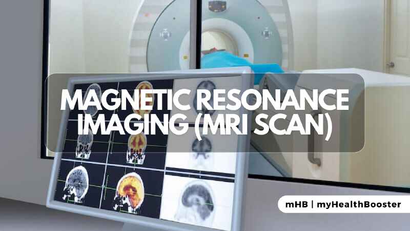
Magnetic Resonance Imaging, or MRI, is a sophisticated medical imaging technique that utilizes a combination of magnetism, radio waves, and computer technology to generate detailed images of various structures within the body. One key advantage of MRI is its ability to produce high-resolution images without exposing patients to potentially harmful x-ray radiation. Moreover, the process itself is painless, offering a non-invasive means of peering into the body’s inner workings.
How It Works
The MRI process involves placing a patient on a movable bed that is then inserted into a tube surrounded by a powerful circular magnet. This magnet generates a robust magnetic field, aligning the protons of hydrogen atoms within the body. When these aligned protons are exposed to radio waves, they emit signals that the MRI scanner detects. A computer processes these signals to create precise and detailed images of the targeted area.
Uses of MRI
MRI serves as an incredibly accurate diagnostic tool, often employed when other imaging tests provide insufficient information. Its applications span a wide range of medical scenarios, from detecting brain trauma, tumors, and aneurysms to evaluating spinal cord integrity after trauma. In addition, MRI is valuable for assessing heart and aorta structures, as well as providing detailed information about abdominal organs, joints, soft tissues, and bones. The information gleaned from an MRI scan can guide medical decisions, including whether surgery is necessary.
Contrast Agents
In certain cases, contrast agents like gadolinium may be introduced to enhance the precision of MRI images, offering a clearer view of specific structures or abnormalities.
Risks and Side Effects
MRI is generally considered safe, with no known side effects. However, it is crucial for patients to inform healthcare providers of any metallic materials within their bodies, as these can distort images. Conditions such as the presence of heart pacemakers or metal implants may limit MRI suitability. Patients may also experience claustrophobia during the procedure, but medical staff are well-trained to assist, and sedatives can be provided when needed.
Preparation and Procedure
Before an MRI scan, metallic objects are removed, and patients may receive sedatives for relaxation. During the procedure, lying still is essential for optimal image accuracy. The MRI machine emits loud clicking noises during scanning, and occasionally, intravenous injections may be administered for contrast imaging.
Results and Interpretation
Following the completion of an MRI scan, a radiologist, a specialized doctor trained in interpreting medical images, analyzes the results. The findings are then compiled into a comprehensive report for the referring healthcare practitioner, who discusses the results with the patient and their family.
Advancements in MRI
Scientists are continually innovating in the field of MRI technology. Newer, smaller, and portable MRI scanners are being developed, with potential applications for detecting infections and tumors in specific body parts. These advancements are currently undergoing testing in medical practice.
Understanding the nuances of MRI empowers individuals, ensuring they approach this diagnostic tool with knowledge and confidence. As technology evolves, the future promises even more refined and accessible MRI capabilities, further enhancing our ability to explore the intricate details of the human body.