A chest X-ray is a powerful diagnostic tool that unveils crucial information about the internal structures of the chest, including the lungs, heart, and bones. In this article, we learn about chest X-rays, examining what they can reveal, the procedure itself, and their significance in medical diagnosis.
Key Points About Chest X-Ray
- Detecting Abnormalities: Chest X-rays are adept at identifying lung and heart irregularities, such as tumors and rib fractures.
- Versatility: While commonly used for lung assessments, chest X-rays can also show abnormalities in the heart, aorta, and thoracic bones.
- Preparation: To ensure accuracy, metallic objects like jewelry are removed to avoid interference with X-ray penetration.
Chest X-Ray Procedure
A chest X-ray involves exposing the chest briefly to radiation to create an image of internal organs. Here’s a breakdown of the procedure:
- Radiation Exposure: The patient wears an X-ray gown, and a small dose of radiation is directed through the chest.
- Tissue Absorption: Different body tissues absorb radiation to varying extents based on their composition, creating shadows on the X-ray.
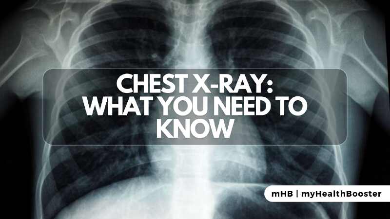
- Shadow Interpretation: White shadows represent dense tissues like bone or the heart, while darker shadows indicate air-filled tissues like the lungs.
Interpretation of Chest X-Ray
Radiologists, physicians specializing in image interpretation, play a pivotal role in analyzing chest X-rays. Modern technology has streamlined this process:
- Digital Advancements: Traditional lightbox methods have been replaced by digital technology, enabling immediate interpretation on computer screens.
- Efficiency: Digitalization facilitates quick analysis, eliminates the risk of lost X-rays, and enhances communication between healthcare professionals.
Where Chest X-Rays Are Performed
Commonly ordered, chest X-rays can be conducted in various settings, including hospitals, emergency rooms, outpatient radiology facilities, and selected doctor’s offices.
Risks and Considerations
While chest X-rays expose patients to minimal radiation, precautions are taken to minimize risks, guided by established standards. Pregnant women are particularly cautious, and protective measures are implemented to shield the fetus from radiation.
Common Reasons for Chest X-Ray
Doctors order chest X-rays for diverse reasons, ranging from respiratory symptoms like shortness of breath to routine check examinations. Pre-operative chest X-rays are also conducted before surgeries to assess heart or lung conditions.
Interpreters of Chest X-Ray
Numerous medical professionals, including radiologists, primary care physicians, and specialists like cardiologists and pulmonologists, are trained to interpret chest X-rays based on their expertise.
Normal and Abnormal Findings
A normal chest X-ray reveals the chest’s size, shape, and key structures. Abnormalities, often in conjunction with clinical data, may include lung conditions (fluid overload, pneumonia), heart abnormalities, and bone-related issues.
Chest X-rays serve as invaluable tools in the diagnostic realm, offering insights into a wide array of conditions affecting the chest. Understanding the procedure, its interpretation, and the significance of findings enhances the collaborative efforts of healthcare professionals in ensuring accurate diagnoses and tailored treatment plans.